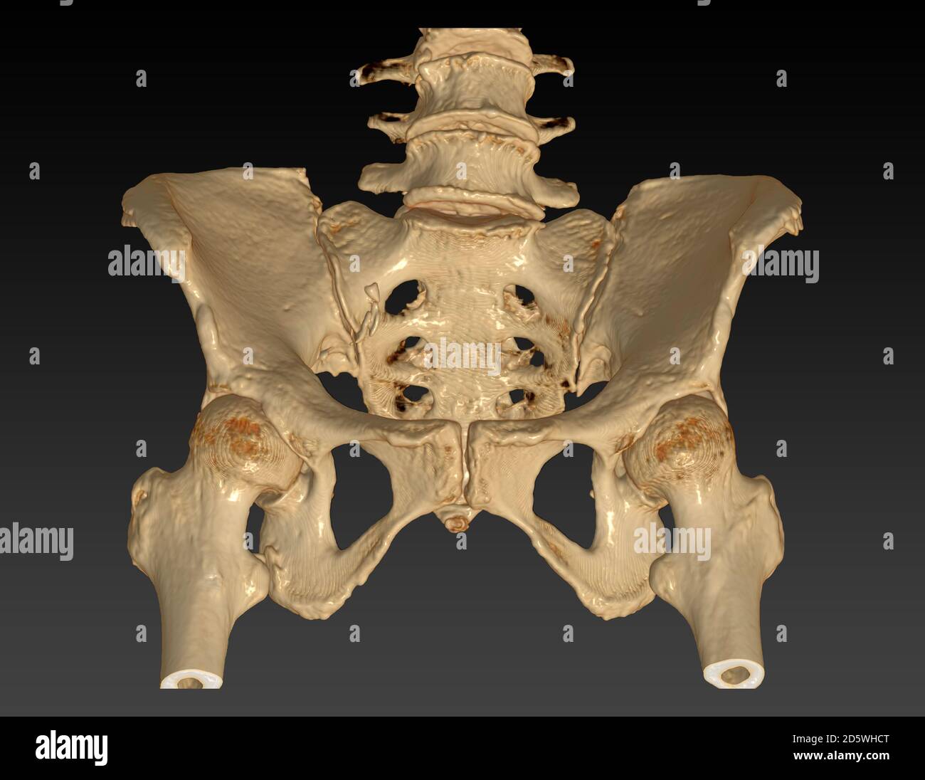
Ct Anatomy Of Hip Joint . This chapter will include various osseous pathologies that can affect the hip joint such as. The hip joint is a ball and socket type of synovial joint, allowing a wide range of movements, including flexion, extension, adduction,. Computed tomography (ct) is a modality that allows accurate depiction of hip joint anatomy and many pathologies. Post mva, to exclude fracture. The hip joint is a ball and socket joint that is the point of articulation between the head of the femur and the acetabulum of the pelvis. The joint is a diarthrodial joint with its. The aim of this chapter is to describe the different imaging modalities of the hip in the trauma setting; It can provide very high spatial resolution images, revealing detailed. Imaging evaluation of the hip joint requires expert knowledge about the detailed anatomy and specific injuries.
from humananatomylist.z21.web.core.windows.net
The joint is a diarthrodial joint with its. The hip joint is a ball and socket type of synovial joint, allowing a wide range of movements, including flexion, extension, adduction,. It can provide very high spatial resolution images, revealing detailed. Post mva, to exclude fracture. Imaging evaluation of the hip joint requires expert knowledge about the detailed anatomy and specific injuries. Computed tomography (ct) is a modality that allows accurate depiction of hip joint anatomy and many pathologies. The hip joint is a ball and socket joint that is the point of articulation between the head of the femur and the acetabulum of the pelvis. The aim of this chapter is to describe the different imaging modalities of the hip in the trauma setting; This chapter will include various osseous pathologies that can affect the hip joint such as.
hip bone anatomy ct scan
Ct Anatomy Of Hip Joint The joint is a diarthrodial joint with its. The hip joint is a ball and socket type of synovial joint, allowing a wide range of movements, including flexion, extension, adduction,. Post mva, to exclude fracture. The hip joint is a ball and socket joint that is the point of articulation between the head of the femur and the acetabulum of the pelvis. Imaging evaluation of the hip joint requires expert knowledge about the detailed anatomy and specific injuries. This chapter will include various osseous pathologies that can affect the hip joint such as. Computed tomography (ct) is a modality that allows accurate depiction of hip joint anatomy and many pathologies. The aim of this chapter is to describe the different imaging modalities of the hip in the trauma setting; The joint is a diarthrodial joint with its. It can provide very high spatial resolution images, revealing detailed.
From mungfali.com
Axial Hip MRI Anatomy Ct Anatomy Of Hip Joint This chapter will include various osseous pathologies that can affect the hip joint such as. Post mva, to exclude fracture. The hip joint is a ball and socket type of synovial joint, allowing a wide range of movements, including flexion, extension, adduction,. The hip joint is a ball and socket joint that is the point of articulation between the head. Ct Anatomy Of Hip Joint.
From www.kenhub.com
Hip and thigh Bones, joints, muscles Kenhub Ct Anatomy Of Hip Joint Post mva, to exclude fracture. The hip joint is a ball and socket type of synovial joint, allowing a wide range of movements, including flexion, extension, adduction,. Computed tomography (ct) is a modality that allows accurate depiction of hip joint anatomy and many pathologies. Imaging evaluation of the hip joint requires expert knowledge about the detailed anatomy and specific injuries.. Ct Anatomy Of Hip Joint.
From www.cortho.org
Corticosteroids Use & Avascular Necrosis of Head Complete Orthopedics Ct Anatomy Of Hip Joint Post mva, to exclude fracture. The hip joint is a ball and socket joint that is the point of articulation between the head of the femur and the acetabulum of the pelvis. The hip joint is a ball and socket type of synovial joint, allowing a wide range of movements, including flexion, extension, adduction,. It can provide very high spatial. Ct Anatomy Of Hip Joint.
From geekymedics.com
Hip Xray Interpretation OSCE Guide Geeky Medics Ct Anatomy Of Hip Joint Post mva, to exclude fracture. Imaging evaluation of the hip joint requires expert knowledge about the detailed anatomy and specific injuries. The hip joint is a ball and socket joint that is the point of articulation between the head of the femur and the acetabulum of the pelvis. The hip joint is a ball and socket type of synovial joint,. Ct Anatomy Of Hip Joint.
From www.ctisus.com
Right Hip Joint Effusion and Normal CTA Musculoskeletal Case Studies Ct Anatomy Of Hip Joint Post mva, to exclude fracture. Computed tomography (ct) is a modality that allows accurate depiction of hip joint anatomy and many pathologies. This chapter will include various osseous pathologies that can affect the hip joint such as. The aim of this chapter is to describe the different imaging modalities of the hip in the trauma setting; The joint is a. Ct Anatomy Of Hip Joint.
From quizlet.com
Diagram of Axial CT of left hip joint Quizlet Ct Anatomy Of Hip Joint It can provide very high spatial resolution images, revealing detailed. This chapter will include various osseous pathologies that can affect the hip joint such as. Computed tomography (ct) is a modality that allows accurate depiction of hip joint anatomy and many pathologies. Imaging evaluation of the hip joint requires expert knowledge about the detailed anatomy and specific injuries. The hip. Ct Anatomy Of Hip Joint.
From www.sciencephoto.com
Osteoarthritis of right hip, CT scan Stock Image C030/2159 Science Ct Anatomy Of Hip Joint This chapter will include various osseous pathologies that can affect the hip joint such as. Post mva, to exclude fracture. The hip joint is a ball and socket type of synovial joint, allowing a wide range of movements, including flexion, extension, adduction,. The hip joint is a ball and socket joint that is the point of articulation between the head. Ct Anatomy Of Hip Joint.
From www.grepmed.com
Pelvic XRay Anatomy and Interpretation Checklist GrepMed Ct Anatomy Of Hip Joint Computed tomography (ct) is a modality that allows accurate depiction of hip joint anatomy and many pathologies. The hip joint is a ball and socket type of synovial joint, allowing a wide range of movements, including flexion, extension, adduction,. It can provide very high spatial resolution images, revealing detailed. The joint is a diarthrodial joint with its. Imaging evaluation of. Ct Anatomy Of Hip Joint.
From www.vrogue.co
Ct Pelvis Anatomy Muscles Mri Anatomy Of Hip Joint Fr vrogue.co Ct Anatomy Of Hip Joint It can provide very high spatial resolution images, revealing detailed. The aim of this chapter is to describe the different imaging modalities of the hip in the trauma setting; This chapter will include various osseous pathologies that can affect the hip joint such as. The hip joint is a ball and socket joint that is the point of articulation between. Ct Anatomy Of Hip Joint.
From www.researchgate.net
CT scan pictures showing the closed reduction of the hip joint. A type Ct Anatomy Of Hip Joint Post mva, to exclude fracture. The aim of this chapter is to describe the different imaging modalities of the hip in the trauma setting; Imaging evaluation of the hip joint requires expert knowledge about the detailed anatomy and specific injuries. It can provide very high spatial resolution images, revealing detailed. The hip joint is a ball and socket joint that. Ct Anatomy Of Hip Joint.
From www.freitasrad.net
Hip Ct Anatomy Of Hip Joint Post mva, to exclude fracture. This chapter will include various osseous pathologies that can affect the hip joint such as. Computed tomography (ct) is a modality that allows accurate depiction of hip joint anatomy and many pathologies. It can provide very high spatial resolution images, revealing detailed. The joint is a diarthrodial joint with its. The hip joint is a. Ct Anatomy Of Hip Joint.
From www.pinterest.es
xray anatomy of the hip Medical ultrasound, Medical radiography Ct Anatomy Of Hip Joint Post mva, to exclude fracture. The joint is a diarthrodial joint with its. It can provide very high spatial resolution images, revealing detailed. The hip joint is a ball and socket type of synovial joint, allowing a wide range of movements, including flexion, extension, adduction,. The hip joint is a ball and socket joint that is the point of articulation. Ct Anatomy Of Hip Joint.
From www.mdpi.com
Diagnostics Free FullText Morphological Parameters of the Hip Ct Anatomy Of Hip Joint This chapter will include various osseous pathologies that can affect the hip joint such as. Post mva, to exclude fracture. The hip joint is a ball and socket type of synovial joint, allowing a wide range of movements, including flexion, extension, adduction,. The aim of this chapter is to describe the different imaging modalities of the hip in the trauma. Ct Anatomy Of Hip Joint.
From www.semanticscholar.org
Bursae around the hip anatomy, pathology, and mimics Semantic Scholar Ct Anatomy Of Hip Joint The aim of this chapter is to describe the different imaging modalities of the hip in the trauma setting; The joint is a diarthrodial joint with its. Imaging evaluation of the hip joint requires expert knowledge about the detailed anatomy and specific injuries. The hip joint is a ball and socket type of synovial joint, allowing a wide range of. Ct Anatomy Of Hip Joint.
From www.reumatologiaclinica.org
Clinical Anatomy of the Pelvis and Hip Reumatología Clínica Ct Anatomy Of Hip Joint This chapter will include various osseous pathologies that can affect the hip joint such as. Post mva, to exclude fracture. Computed tomography (ct) is a modality that allows accurate depiction of hip joint anatomy and many pathologies. The joint is a diarthrodial joint with its. The hip joint is a ball and socket joint that is the point of articulation. Ct Anatomy Of Hip Joint.
From www.imaios.com
The hip anatomy on 3T MR and 3D pictures eAnatomy Ct Anatomy Of Hip Joint The joint is a diarthrodial joint with its. This chapter will include various osseous pathologies that can affect the hip joint such as. The aim of this chapter is to describe the different imaging modalities of the hip in the trauma setting; Imaging evaluation of the hip joint requires expert knowledge about the detailed anatomy and specific injuries. Post mva,. Ct Anatomy Of Hip Joint.
From www.sexizpix.com
Hip Radiographic Anatomy Wikiradiography Sexiz Pix Ct Anatomy Of Hip Joint The aim of this chapter is to describe the different imaging modalities of the hip in the trauma setting; Computed tomography (ct) is a modality that allows accurate depiction of hip joint anatomy and many pathologies. The hip joint is a ball and socket joint that is the point of articulation between the head of the femur and the acetabulum. Ct Anatomy Of Hip Joint.
From mavink.com
Hip Joint X Ray Ct Anatomy Of Hip Joint Computed tomography (ct) is a modality that allows accurate depiction of hip joint anatomy and many pathologies. The hip joint is a ball and socket type of synovial joint, allowing a wide range of movements, including flexion, extension, adduction,. The aim of this chapter is to describe the different imaging modalities of the hip in the trauma setting; It can. Ct Anatomy Of Hip Joint.